Perioperative Interactive Education
The Perioperative Interactive Education (PIE) team in the Department of Anesthesia and Pain Management at Toronto General Hospital is developing a wide variety of online teaching aids to enhance the teaching of perioperative medicine. These resources aids are intended to be used both by medical educators in small group sessions or lectures and by trainees for self-study. They are all freely available on the Web to educators and trainees around the world. The PIE Web site (http://pie.med.utoronto.ca) describes all the projects under development and the team members who are involved in this initiative. The projects are available in four Web sites:
 | ||
 | ||
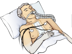
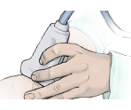
 Cardiac AnesthesiaThis Web site contains teaching material relevant to perioperative care. It contains an interactive module illustrating the movement of water between the body fluid compartments in fluid management, and a module describing the factors involved in the control of blood pressure and cardiac output (Starling’s Law). Patient simulations of postoperative cardiac patients and weaning patients from cardiopulmonary bypass are under development. | ||
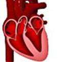
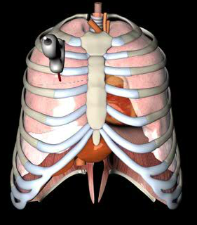
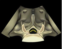
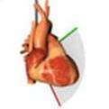
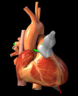
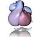 Cardiac Embryologyhttp://pie.med.utoronto.ca/htbg This Web site illustrates the embryologic development of the heart from the cardiac crest to the separation of the outflow tracts using a 3D model which can be rotated horizontally to view the model from any angle. A time line can be dragged to show the growth and development between each of the stage. | ||
 | ||
 | ||
 | ||
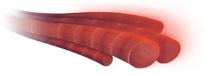
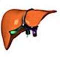 Virtual Liverhttp://pie.med.utoronto.ca/vliver This Web site helps trainees learning to interpret the anatomical structures in CTs of the liver in surgical planning. We have created a 3D model of a liver and its internal structure of arteries, veins, portal and biliary systems. The model can be made transparent to view the internal structures, and the model rotated either horizontally or vertically to view it from any angle. The structures can be labeled, and the corresponding structures in the CT can be seen alongside the 3D model. The Cuinaud segments and standard surgical resections of the liver can also be viewed with different levels of transparency to see the internal structure related to these segments. | ||
 | ||
 | ||
 |